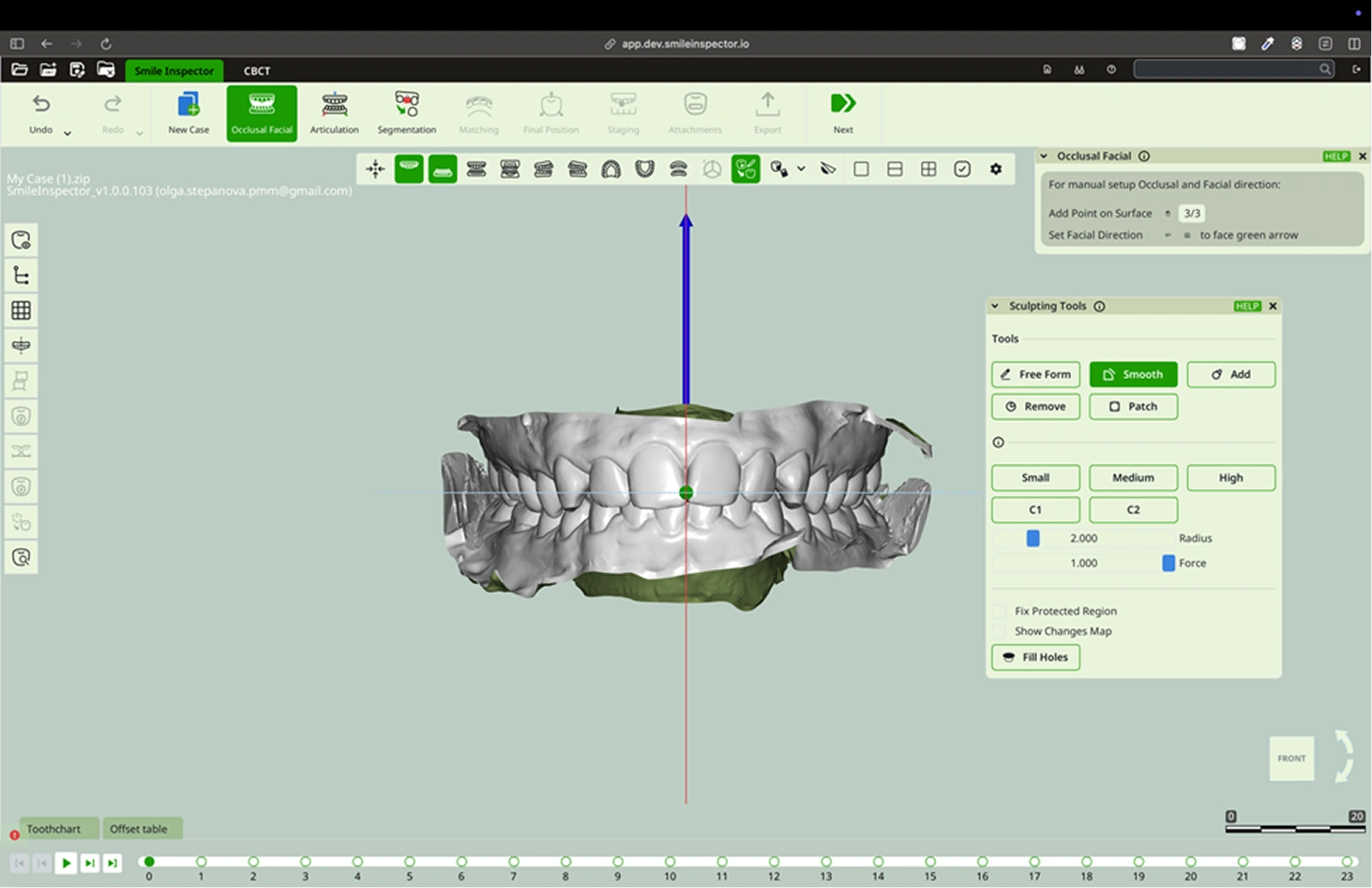
What: Modify mesh surfaces manually.
How:
- Freeform is used to modify the tooth surface according to the model’s position. Position it in a side view to create indentations or increase the tooth surface. Position it in a front view to adjust the tooth size, tooth angles, or the shape of the cervical margins.
- Smooth is used to soften surfaces. If you create a groove using the Remove tool, you can smooth it with Smooth.
- To add material in indentations or recreate a missing part of the tooth, use the Add tool.
- With the Remove tool, you can remove material, create indentations, or carve grooves to define the cervical margins in scans where it is needed.
- Patch quickly fill surfaces, remove material, and repair damaged areas of the scan.
- The Small, Medium, and High buttons activate predefined size and strength settings for the tools.
- The C1 and C2 buttons are options to save size and strength settings for each tool. Choose the desired values using the Radius and Force sliders, then press Save to store the settings. Use the Cancel button to exit the save mode.
- Check the Fix Protected Region box to protect an area from being modified while sculpting.
- To view the modified dental area, enable Show Changes Map.
- Fill Holes fills all the gaps in the scan.
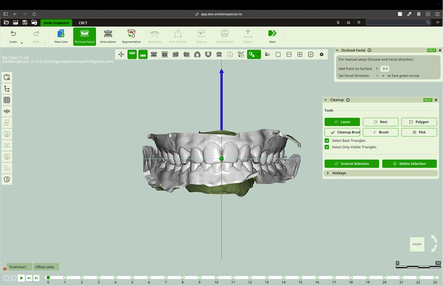
Cleanup Tools
What: Delete unwanted mesh parts.
How:
- Use selection tools:
1.1. Lasso: Select the entire surface to be modify within a stroke drawn by the cursor.
1.2. Rect: Marks the surface in a straight, square shape.
1.3 Polygon: Marks the surface using the vertices of a polygon.
1.4 Clean Up Brush: Selects the opposite area, expanding it based on the drawn shape.
1.5 Brush: Selects only the area painted under the brush.
1.6 Pick: Selects a very small area or a single triangle to modify..
- Mark and Delete Selection.
- Use Inverse Selection if needed to select opposite area of the one marked.
Section
What: Show cross section of objects by user selected plane.
How:
Draw a line across the plane and press Cut Objects.
View Ports

There are three types of viewports: a single window, two horizontal windows, and four windows.
There are three types of viewports: a single window, two horizontal windows, and four windows.
Left Toolbar
Visibility: Within this option, features such as Jaws, Scans, Gingiva, Gingiva Scan, Crowns, Teeth, Arch, and Individual Teeth can be enabled or disabled.
Grid: Shows or hides the grid for leveling.
Midpline: Show Midplane Graphics.
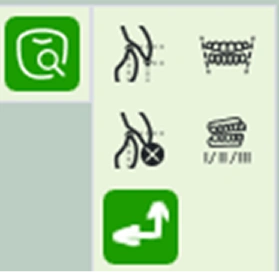
Occlusogram: Allows visualization of the occlusal contact points between arches.
Movement Color: Show Teeth coloring according to their movement.
IP Contacts: Displays the interdental contacts. With options such as:
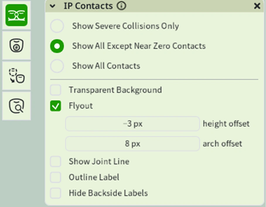
- Show sever collisions only
- Show all except near zero contacts.
- Show all contacts.
- Transparent background.
- Show Joint Line.
- Outline Label.
- Hide back side labels.
Teeth numeration: Displays the tooth numbers.
Initial Teeth position: Show or hide teeth on initial stage.
Diagnostics:
- Overbite Overjet: Calculates the vertical (overbite) and horizontal (overjet) overlap of the arches, Includes the mode options Midline Aligned and Arch Follow. Enter the positioning measurement in the bar.
- Bolton Analysis: Analysis to determine quality of inter arch relationship.
- Alveolar Dsicrepancy: Analysis to clasify crowding or spacing for each jaw.
- Bite clasification: Determine bite clase l, ll or lll.
- Anatomical Basis: Show or hide anatomical basis.
CBCT
What: Performs the diagnosis of the intraoral scan using the CBCT scan.
How:
Change ISO: Select what ISO surface will represent the voxel object on the screen, and choose the method construct the ISO Surface.
Voxels Slice: Show volume slice as image.
Set Active Voxel Box: Set area for work on boxes.
Align IO Scans to CBCT: Align intraoral scans to the CBCT scan.
Bones extraction: Extract skull bones from CBCT Scans.
Contact and Support
For questions or assistance, please contact:
support@smileinspector.io
This manual is subject to updates as features evolve. Please refer to the latest version at smileinspector.io/support.